X-rays briefly. Application of X-ray radiation in medicine. Receiving X-rays
X-ray radiation (synonym X-rays) is with a wide range of wavelengths (from 8·10 -6 to 10 -12 cm). X-ray radiation occurs when charged particles, most often electrons, are decelerated in the electric field of atoms of a substance. The quanta formed in this case have different energies and form a continuous spectrum. The maximum energy of quanta in such a spectrum is equal to the energy of incident electrons. B (cm.) maximum energy quanta x-ray radiation, expressed in kiloelectron-volts, is numerically equal to the magnitude of the voltage applied to the tube, expressed in kilovolts. When X-rays pass through a substance, they interact with the electrons of its atoms. For X-ray quanta with energies up to 100 keV, the most characteristic type of interaction is the photoelectric effect. As a result of such interaction, the energy of the quantum is completely spent on tearing the electron out of the atomic shell and imparting kinetic energy to it. As the energy of an X-ray quantum increases, the probability of the photoelectric effect decreases and the process of scattering of quantums by free electrons - the so-called Compton effect - becomes predominant. As a result of such interaction, a secondary electron is also formed and, in addition, a quantum is emitted with an energy lower than the energy of the primary quantum. If the energy of the X-ray quantum exceeds one megaelectron-volt, the so-called pairing effect can occur, in which an electron and a positron are formed (see). Consequently, when passing through a substance, the energy of X-ray radiation decreases, i.e., its intensity decreases. Since absorption of low-energy quanta occurs with a greater probability, the X-ray radiation is enriched with higher-energy quanta. This property of X-ray radiation is used to increase the average energy of quanta, i.e., to increase its hardness. An increase in the hardness of X-ray radiation is achieved using special filters (see). X-ray radiation is used for x-ray diagnostics (see) and (see). See also Ionizing radiation.
X-ray radiation (synonym: x-rays, x-rays) is quantum electromagnetic radiation with a wavelength from 250 to 0.025 A (or energy quanta from 5·10 -2 to 5·10 2 keV). In 1895 it was discovered by V.K. Roentgen. Spectral region adjacent to X-ray radiation electromagnetic radiation, whose energy quanta exceed 500 keV, are called gamma radiation (see); radiation whose energy quanta are below 0.05 kev constitutes ultraviolet radiation (see).
Thus, representing a relatively small part of the vast spectrum of electromagnetic radiation, which includes both radio waves and visible light, X-ray radiation, like any electromagnetic radiation, propagates at the speed of light (in a vacuum of about 300 thousand km/sec) and is characterized by a wavelength λ ( the distance over which radiation travels in one oscillation period). X-ray radiation also has a number of other wave properties (refraction, interference, diffraction), but they are much more difficult to observe than longer wavelength radiation: visible light, radio waves.
X-ray spectra: a1 - continuous bremsstrahlung spectrum at 310 kV; a - continuous brake spectrum at 250 kV, a1 - spectrum filtered with 1 mm Cu, a2 - spectrum filtered with 2 mm Cu, b - K-series tungsten lines.
To generate X-ray radiation, X-ray tubes (see) are used, in which radiation occurs when fast electrons interact with atoms of the anode substance. There are two types of X-ray radiation: bremsstrahlung and characteristic. Bremsstrahlung X-rays have a continuous spectrum, similar to ordinary white light. The intensity distribution depending on the wavelength (Fig.) is represented by a curve with a maximum; towards long waves the curve falls flatly, and towards short waves it falls steeply and ends at a certain wavelength (λ0), called the short-wave boundary of the continuous spectrum. The value of λ0 is inversely proportional to the voltage on the tube. Bremsstrahlung occurs when fast electrons interact with atomic nuclei. The intensity of bremsstrahlung is directly proportional to the strength of the anode current, the square of the voltage across the tube and the atomic number (Z) of the anode substance.
If the energy of the electrons accelerated in the X-ray tube exceeds the value critical for the anode substance (this energy is determined by the voltage Vcr critical for this substance on the tube), then characteristic radiation occurs. The characteristic spectrum is lined; its spectral lines form series, designated by the letters K, L, M, N.
Series K is the shortest wavelength, series L is longer wavelength, series M and N are observed only in heavy elements(Vcr of tungsten for the K-series - 69.3 kV, for the L-series - 12.1 kV). Characteristic radiation arises as follows. Fast electrons knock atomic electrons out of their inner shells. The atom is excited and then returns to the ground state. In this case, electrons from the outer, less bound shells fill the vacated inner shells places, and photons of characteristic radiation are emitted with an energy equal to the difference in the energies of the atom in the excited and ground states. This difference (and therefore the photon energy) has a certain value characteristic of each element. This phenomenon underlies X-ray spectral analysis of elements. The figure shows the line spectrum of tungsten against the background of a continuous spectrum of bremsstrahlung.
The energy of electrons accelerated in the X-ray tube is converted almost entirely into thermal energy (the anode becomes very hot), only a small part (about 1% at a voltage close to 100 kV) is converted into bremsstrahlung energy.
The use of X-rays in medicine is based on the laws of absorption of X-rays by matter. X-ray absorption is completely independent of optical properties absorbent substances. Colorless and transparent lead glass, used to protect personnel in x-ray rooms, almost completely absorbs x-rays. In contrast, a sheet of paper that is not transparent to light does not attenuate x-rays.
The intensity of a homogeneous (i.e., a certain wavelength) X-ray beam passing through an absorber layer decreases according to the exponential law (e-x), where e is the base of natural logarithms (2.718), and the exponent x is equal to the product of the mass attenuation coefficient (μ /p) cm 2 /g per thickness of the absorber in g/cm 2 (here p is the density of the substance in g/cm 3). The attenuation of X-ray radiation occurs due to both scattering and absorption. Accordingly, the mass attenuation coefficient is the sum of the mass absorption and scattering coefficients. The mass absorption coefficient increases sharply with increasing atomic number (Z) of the absorber (proportional to Z3 or Z5) and with increasing wavelength (proportional to λ3). This dependence on wavelength is observed within the absorption bands, at the boundaries of which the coefficient exhibits jumps.
The mass scattering coefficient increases with increasing atomic number of the substance. At λ≥0.3Å the scattering coefficient does not depend on the wavelength, at λ<0,ЗÅ он уменьшается с уменьшением λ.
A decrease in the absorption and scattering coefficients with decreasing wavelength causes an increase in the penetrating power of X-ray radiation. The mass absorption coefficient for bone [uptake is mainly due to Ca 3 (PO 4) 2 ] is almost 70 times greater than for soft tissue, where uptake is mainly due to water. This explains why the shadow of bones stands out so sharply against the background of soft tissue on radiographs.
The propagation of a non-uniform X-ray beam through any medium, along with a decrease in intensity, is accompanied by a change in the spectral composition and a change in the quality of the radiation: the long-wave part of the spectrum is absorbed to a greater extent than the short-wave part, the radiation becomes more homogeneous. Filtering out the long-wave part of the spectrum allows, during X-ray therapy of lesions located deep in the human body, to improve the ratio between deep and surface doses (see X-ray filters). To characterize the quality of an inhomogeneous beam of X-rays, the concept of “half-attenuation layer (L)” is used - a layer of substance that attenuates the radiation by half. The thickness of this layer depends on the voltage on the tube, the thickness and material of the filter. To measure half-attenuation layers, cellophane (up to 12 keV energy), aluminum (20-100 keV), copper (60-300 keV), lead and copper (>300 keV) are used. For X-rays generated at voltages of 80-120 kV, 1 mm of copper is equivalent in filtering capacity to 26 mm of aluminum, 1 mm of lead is equivalent to 50.9 mm of aluminum.
The absorption and scattering of X-ray radiation is due to its corpuscular properties; X-ray radiation interacts with atoms as a stream of corpuscles (particles) - photons, each of which has a certain energy (inversely proportional to the wavelength of X-ray radiation). The energy range of X-ray photons is 0.05-500 keV.
The absorption of X-ray radiation is due to the photoelectric effect: the absorption of a photon by the electron shell is accompanied by the ejection of an electron. The atom is excited and, returning to the ground state, emits characteristic radiation. The emitted photoelectron carries away all the energy of the photon (minus the binding energy of the electron in the atom).
X-ray scattering is caused by electrons in the scattering medium. A distinction is made between classical scattering (the wavelength of the radiation does not change, but the direction of propagation changes) and scattering with a change in wavelength - the Compton effect (the wavelength of the scattered radiation is greater than that of the incident radiation). In the latter case, the photon behaves like a moving ball, and the scattering of photons occurs, according to Comton’s figurative expression, like playing billiards with photons and electrons: colliding with an electron, the photon transfers part of its energy to it and is scattered, having less energy (accordingly, the wavelength of the scattered radiation increases), an electron flies out of the atom with recoil energy (these electrons are called Compton electrons, or recoil electrons). Absorption of X-ray energy occurs during the formation of secondary electrons (Compton and photoelectrons) and the transfer of energy to them. The energy of X-ray radiation transferred to a unit mass of a substance determines the absorbed dose of X-ray radiation. The unit of this dose 1 rad corresponds to 100 erg/g. Due to the absorbed energy, a number of secondary processes occur in the absorber substance, which are important for X-ray dosimetry, since it is on them that the methods for measuring X-ray radiation are based. (see Dosimetry).
All gases and many liquids, semiconductors and dielectrics increase electrical conductivity when exposed to X-rays. Conductivity is detected by the best insulating materials: paraffin, mica, rubber, amber. The change in conductivity is caused by ionization of the medium, i.e., the separation of neutral molecules into positive and negative ions (ionization is produced by secondary electrons). Ionization in air is used to determine X-ray exposure dose (dose in air), which is measured in roentgens (see Ionizing Radiation Doses). At a dose of 1 r, the absorbed dose in air is 0.88 rad.
Under the influence of X-ray radiation, as a result of the excitation of molecules of a substance (and during the recombination of ions), in many cases a visible glow of the substance is excited. At high intensities of X-ray radiation, a visible glow is observed in air, paper, paraffin, etc. (with the exception of metals). The highest yield of visible luminescence is provided by crystalline phosphors such as Zn·CdS·Ag-phosphorus and others used for fluoroscopy screens.
Under the influence of X-ray radiation, various chemical processes: decomposition of silver halide compounds (photographic effect used in radiography), decomposition of water and aqueous solutions of hydrogen peroxide, change in the properties of celluloid (turbidity and release of camphor), paraffin (turbidity and bleaching).
As a result of complete conversion, all the energy absorbed by the chemically inert substance, the x-ray radiation, is converted into heat. Measuring very small amounts of heat requires highly sensitive methods, but is the main method for absolute measurements of X-ray radiation.
Secondary biological effects from exposure to x-ray radiation are the basis of medical x-ray therapy (see). X-ray radiation, whose quanta are 6-16 keV (effective wavelengths from 2 to 5 Å), is almost completely absorbed by the skin tissue of the human body; these are called boundary rays, or sometimes Bucca's rays (see Bucca's rays). For deep X-ray therapy, hard filtered radiation with effective energy quanta from 100 to 300 keV is used.
The biological effect of X-ray radiation should be taken into account not only during X-ray therapy, but also during X-ray diagnostics, as well as in all other cases of contact with X-ray radiation that require the use of radiation protection (see).
X-rays were discovered by accident in 1895 by the famous German physicist Wilhelm Roentgen. He studied cathode rays in a low-pressure gas-discharge tube at high voltage between its electrodes. Despite the fact that the tube was in a black box, Roentgen noticed that a fluorescent screen, which happened to be nearby, glowed every time the tube was in use. The tube turned out to be a source of radiation that could penetrate paper, wood, glass and even a one and a half centimeter thick aluminum plate.
X-ray determined that the gas-discharge tube was a source of a new type of invisible radiation with great penetrating power. The scientist could not determine whether this radiation was a stream of particles or waves, and he decided to give it the name X-rays. They were later called X-rays
It is now known that X-rays are a type of electromagnetic radiation that has a shorter wavelength than ultraviolet electromagnetic waves. The wavelength of X rays ranges from 70 nm up to 10 -5 nm. The shorter the wavelength of X-rays, the greater the energy of their photons and the greater their penetrating power. X-rays with a relatively long wavelength (more than 10 nm), are called soft. Wavelength 1 - 10 nm characterizes hard X-rays. They have enormous penetrating power.
Receiving X-rays
X-rays are produced when fast electrons, or cathode rays, collide with the walls or anode of a low-pressure gas discharge tube. A modern X-ray tube is a evacuated glass cylinder with a cathode and anode located in it. The potential difference between the cathode and anode (anti-cathode) reaches several hundred kilovolts. The cathode is a tungsten filament heated by electric current. This causes the cathode to emit electrons as a result of thermionic emission. The electrons are accelerated by the electric field in the X-ray tube. Since there is a very small number of gas molecules in the tube, the electrons practically do not lose their energy on the way to the anode. They reach the anode at a very high speed.
X-rays are produced whenever electrons moving at high speed are slowed down by the anode material. Most of the electrons' energy is dissipated as heat. Therefore, the anode must be artificially cooled. The anode in the X-ray tube must be made of a metal that has a high melting point, such as tungsten.
The part of the energy that is not dissipated in the form of heat is converted into the energy of electromagnetic waves (X-rays). Thus, X-rays are the result of electron bombardment of the anode substance. There are two types of X-rays: bremsstrahlung and characteristic.
Bremsstrahlung X-rays
Bremsstrahlung X-rays occur when electrons moving at high speed are decelerated. electric fields atoms of the anode. The conditions for stopping individual electrons are not the same. As a result, various parts of their kinetic energy are converted into X-ray energy.
The spectrum of X-ray bremsstrahlung does not depend on the nature of the anode substance. As is known, the energy of X-ray photons determines their frequency and wavelength. Therefore, X-ray bremsstrahlung is not monochromatic. It is characterized by a variety of wavelengths that can be represented continuous (continuous) spectrum.
X-rays cannot have an energy greater than the kinetic energy of the electrons that form them. The shortest wavelength of X-ray radiation corresponds to the maximum kinetic energy of decelerating electrons. The greater the potential difference in the X-ray tube, the shorter the wavelengths of X-ray radiation can be obtained.
Characteristic X-ray radiation
The characteristic X-ray radiation is not continuous, but line spectrum. This type of radiation occurs when a fast electron, reaching the anode, penetrates the inner orbitals of atoms and knocks out one of their electrons. As a result, a free space appears that can be filled by another electron descending from one of the upper atomic orbitals. This transition of an electron from a higher to a lower energy level produces x-rays of a specific discrete wavelength. Therefore, the characteristic X-ray radiation has line spectrum. The frequency of the characteristic radiation lines completely depends on the structure of the electron orbitals of the anode atoms.
The spectrum lines of the characteristic radiation of different chemical elements have the same appearance, since the structure of their internal electron orbitals is identical. But their wavelength and frequency are due to energy differences between the internal orbitals of heavy and light atoms.
The frequency of the lines in the spectrum of characteristic X-ray radiation changes in accordance with the atomic number of the metal and is determined by the Moseley equation: v 1/2 = A(Z-B), Where Z- atomic number chemical element, A And B- constants.
Primary physical mechanisms of interaction of X-ray radiation with matter
The primary interaction between X-rays and matter is characterized by three mechanisms:
1. Coherent scattering. This form of interaction occurs when the X-ray photons have less energy than the binding energy of the electrons to the atomic nucleus. In this case, the photon energy is not sufficient to release electrons from the atoms of the substance. The photon is not absorbed by the atom, but changes the direction of propagation. In this case, the wavelength of X-ray radiation remains unchanged.
2. Photoelectric effect (photoelectric effect). When an X-ray photon reaches an atom of a substance, it can knock out one of the electrons. This occurs if the photon energy exceeds the binding energy of the electron with the nucleus. In this case, the photon is absorbed and the electron is released from the atom. If a photon carries more energy than is needed to release an electron, it will transfer the remaining energy to the released electron in the form of kinetic energy. This phenomenon, called the photoelectric effect, occurs when relatively low-energy X-rays are absorbed.
An atom that loses one of its electrons becomes a positive ion. The lifetime of free electrons is very short. They are absorbed by neutral atoms, which turn into negative ions. The result of the photoelectric effect is intense ionization of the substance.
If the energy of the X-ray photon is less than the ionization energy of the atoms, then the atoms go into an excited state, but are not ionized.
3. Incoherent scattering (Compton effect). This effect was discovered by the American physicist Compton. It occurs when a substance absorbs X-rays of short wavelength. The photon energy of such X-rays is always greater than the ionization energy of the atoms of the substance. The Compton effect results from the interaction of a high-energy X-ray photon with one of the electrons in the outer shell of an atom, which has a relatively weak connection with the atomic nucleus.
A high-energy photon transfers some of its energy to the electron. The excited electron is released from the atom. The remaining energy from the original photon is emitted as an x-ray photon of longer wavelength at some angle to the direction of motion of the original photon. The secondary photon can ionize another atom, etc. These changes in the direction and wavelength of X-rays are known as the Compton effect.
Some effects of interaction of X-rays with matter
As mentioned above, X-rays are capable of exciting atoms and molecules of matter. This may cause fluorescence certain substances(for example, zinc sulfate). If a parallel beam of X-rays is directed at opaque objects, you can observe how the rays pass through the object by placing a screen covered with a fluorescent substance.
The fluorescent screen can be replaced with photographic film. X-rays have the same effect on photographic emulsion as light. Both methods are used in practical medicine.
Another important effect of X-rays is their ionizing ability. This depends on their wavelength and energy. This effect provides a method for measuring the intensity of x-rays. When X-rays pass through an ionization chamber, electricity, the magnitude of which is proportional to the intensity of X-ray radiation.
Absorption of X-rays by matter
As X-rays pass through matter, their energy decreases due to absorption and scattering. The attenuation of the intensity of a parallel beam of X-rays passing through a substance is determined by Bouguer’s law: I = I0 e -μd, Where I 0- initial intensity of X-ray radiation; I- intensity of X-rays passing through the layer of matter, d- absorbent layer thickness , μ - linear attenuation coefficient. It is equal to the sum of two quantities: t- linear absorption coefficient and σ - linear dissipation coefficient: μ = τ+ σ
Experiments have revealed that the linear absorption coefficient depends on the atomic number of the substance and the wavelength of the X-rays:
τ = kρZ 3 λ 3, Where k- coefficient of direct proportionality, ρ - density of the substance, Z- atomic number of the element, λ - wavelength of x-rays.
The dependence on Z is very important from a practical point of view. For example, the absorption coefficient of bones, which are composed of calcium phosphate, is almost 150 times higher than that of soft tissue ( Z=20 for calcium and Z=15 for phosphorus). When X-rays pass through the human body, bones stand out clearly against the background of muscles, connective tissue, etc.
It is known that the digestive organs have the same absorption coefficient as other soft tissues. But the shadow of the esophagus, stomach and intestines can be distinguished if the patient takes a contrast agent - barium sulfate ( Z= 56 for barium). Barium sulfate is very opaque to x-rays and is often used for x-ray examination of the gastrointestinal tract. Certain opaque mixtures are injected into the bloodstream in order to examine the condition of blood vessels, kidneys, etc. In this case, iodine, whose atomic number is 53, is used as a contrast agent.
Dependence of X-ray absorption on Z also used to protect against the possible harmful effects of x-rays. Lead is used for this purpose, the amount Z for which it is equal to 82.
Application of X-rays in medicine
The reason for the use of x-rays in diagnostics was their high penetrating ability, one of the main properties of x-ray radiation. In the early days after its discovery, X-rays were used mostly to examine bone fractures and determine the location of foreign bodies (such as bullets) in the human body. Currently, several diagnostic methods using x-rays (x-ray diagnostics) are used.
X-ray . An X-ray device consists of an X-ray source (X-ray tube) and a fluorescent screen. After X-rays pass through the patient's body, the doctor observes a shadow image of him. A lead window should be installed between the screen and the physician's eyes to protect the physician from the harmful effects of X-rays. This method makes it possible to study the functional state of certain organs. For example, the doctor can directly observe the movements of the lungs and the passage of the contrast agent through the gastrointestinal tract. The disadvantages of this method are insufficient contrast images and relatively large doses of radiation received by the patient during the procedure.
Fluorography . This method consists of taking a photograph of a part of the patient's body. Typically used for preliminary examination of the condition internal organs patients using low doses of X-ray radiation.
Radiography. (X-ray radiography). This is a research method using x-rays in which an image is recorded on photographic film. Photographs are usually taken in two perpendicular planes. This method has some advantages. X-ray photographs contain more detail than a fluorescent screen and are therefore more informative. They can be saved for further analysis. The total radiation dose is less than that used in fluoroscopy.
Computed X-ray tomography . Equipped with computer technology, an axial tomography scanner is the most modern X-ray diagnostic device that allows you to obtain a clear image of any part of the human body, including soft tissues of organs.
The first generation of computed tomography (CT) scanners include a special X-ray tube that is attached to a cylindrical frame. A thin beam of X-rays is directed at the patient. Two X-ray detectors are attached to opposite side frames The patient is in the center of the frame, which can rotate 180° around his body.
An X-ray beam passes through a stationary object. The detectors obtain and record the absorption values of various tissues. Recordings are made 160 times while the X-ray tube moves linearly along the scanned plane. Then the frame is rotated 1 0 and the procedure is repeated. Recording continues until the frame rotates 180 0 . Each detector records 28,800 frames (180x160) during the study. The information is processed by a computer, and an image of the selected layer is formed using a special computer program.
The second generation of CT uses several X-ray beams and up to 30 X-ray detectors. This makes it possible to speed up the research process up to 18 seconds.
The third generation of CT uses a new principle. A wide fan-shaped beam of X-rays covers the object under study, and the X-ray radiation passing through the body is recorded by several hundred detectors. The time required for research is reduced to 5-6 seconds.
CT has many advantages over earlier x-ray diagnostic methods. It is characterized high resolution, which makes it possible to distinguish subtle changes in soft tissues. CT allows you to detect pathological processes that cannot be detected by other methods. In addition, the use of CT makes it possible to reduce the dose of X-ray radiation received by patients during the diagnostic process.
The discovery and merits in the study of the basic properties of X-rays rightfully belong to the German scientist Wilhelm Conrad Roentgen. Amazing Properties The X-rays he discovered immediately received a huge resonance in the scientific world. Although back then, back in 1895, the scientist could hardly have imagined what benefits, and sometimes harm, X-ray radiation could bring.
Let's find out in this article how this type of radiation affects human health.
What is X-ray radiation

The first question that interested the researcher was what is X-ray radiation? A series of experiments made it possible to verify that this is electromagnetic radiation with a wavelength of 10 -8 cm, occupying an intermediate position between ultraviolet and gamma radiation.
Applications of X-rays
All of these aspects of the destructive effects of the mysterious X-rays do not at all exclude surprisingly extensive aspects of their application. Where is X-ray radiation used?
- Study of the structure of molecules and crystals.
- X-ray flaw detection (in industry, detection of defects in products).
- Methods of medical research and therapy.
The most important applications of X-rays are made possible by the very short wavelengths of these waves and their unique properties.
Since we are interested in the effect of X-ray radiation on people who encounter it only during a medical examination or treatment, then we will further consider only this area of application of X-rays.
Application of X-rays in medicine
 Despite the special significance of his discovery, Roentgen did not take out a patent for its use, making it an invaluable gift for all humanity. Already in the First World War, X-ray machines began to be used, which made it possible to quickly and accurately diagnose the wounded. Now we can distinguish two main areas of application of X-rays in medicine:
- X-ray diagnostics;
- X-ray therapy.
X-ray diagnostics
X-ray diagnostics is used in various ways:

Let's look at the differences between these methods.

All of these diagnostic methods are based on the ability of X-rays to illuminate photographic film and on their different permeability to tissues and the bone skeleton.
X-ray therapy
The ability of X-rays to have a biological effect on tissue is used in medicine to treat tumors. The ionizing effect of this radiation is most actively manifested in its effect on rapidly dividing cells, which are the cells of malignant tumors.
However, you should also be aware of the side effects that inevitably accompany x-ray therapy. The fact is that cells of the hematopoietic, endocrine, and immune systems also rapidly divide. Negative effects on them give rise to signs of radiation sickness.
The effect of X-ray radiation on humans
Soon after the remarkable discovery of X-rays, it was discovered that X-rays had an effect on humans.

These data were obtained from experiments on experimental animals, however, geneticists suggest that similar consequences can extend to the human body.
Studying the effects of X-ray exposure has made it possible to develop international standards for permissible radiation doses.
X-ray doses during X-ray diagnostics
After visiting the X-ray room, many patients feel worried about how the received dose of radiation will affect their health?

The dose of total body radiation depends on the nature of the procedure performed. For convenience, we will compare the dose received with natural radiation, which accompanies a person throughout his life.
- X-ray: chest - the received radiation dose is equivalent to 10 days of background radiation; upper stomach and small intestine - 3 years.
- Computed tomography of the abdominal and pelvic organs, as well as the whole body - 3 years.
- Mammography - 3 months.
- X-rays of the extremities are practically harmless.
- As for dental x-rays, the radiation dose is minimal, since the patient is exposed to a narrow beam of x-rays with a short radiation duration.
These radiation doses meet acceptable standards, but if the patient experiences anxiety before undergoing an x-ray, he has the right to request a special protective apron.
Exposure to X-rays in pregnant women
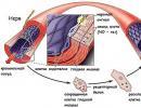
Every person is forced to undergo X-ray examinations more than once. But there is a rule - this diagnostic method cannot be prescribed to pregnant women. The developing embryo is extremely vulnerable. X-rays can cause chromosome abnormalities and, as a result, the birth of children with developmental defects. The most vulnerable period in this regard is pregnancy up to 16 weeks. Moreover, X-rays of the spine, pelvic and abdominal areas are most dangerous for the unborn baby.
 Knowing about the harmful effects of X-ray radiation on pregnancy, doctors in every possible way avoid using it during this important period in a woman’s life.
However, there are side sources of X-ray radiation:
- electron microscopes;
- picture tubes of color TVs, etc.
Expectant mothers should be aware of the danger posed by them.
X-ray diagnostics are not dangerous for nursing mothers.
What to do after an X-ray
To avoid even minimal effects from X-ray exposure, you can take some simple steps:
- after an x-ray, drink a glass of milk - it removes small doses of radiation;
- It’s very helpful to take a glass of dry wine or grape juice;
- For some time after the procedure, it is useful to increase the proportion of foods with a high iodine content (seafood).
But, no medical procedures or special measures are required to remove radiation after an x-ray!
Despite the undoubtedly serious consequences of exposure to X-rays, their danger during medical examinations should not be overestimated - they are carried out only on certain areas of the body and very quickly. The benefits from them many times exceed the risk of this procedure for the human body.
In 1895, the German physicist W. Roentgen discovered a new, previously unknown type of electromagnetic radiation, which was named X-ray in honor of its discoverer. V. Roentgen became the author of his discovery at the age of 50, holding the post of rector of the University of Würzburg and having a reputation as one of the best experimenters of his time. One of the first to find technical application for the discovery of X-ray was the American Edison. He created a convenient demonstration apparatus and already in May 1896 organized an X-ray exhibition in New York, where visitors could examine their own hand on a luminous screen. After Edison's assistant died from severe burns he received during constant demonstrations, the inventor stopped further experiments with X-rays.
X-ray radiation began to be used in medicine due to its high penetrating ability. Initially, X-rays were used to examine bone fractures and determine the location of foreign bodies in the human body. Currently, there are several methods based on X-ray radiation. But these methods have their drawbacks: radiation can cause deep damage to the skin. The ulcers that appeared often turned into cancer. In many cases, fingers or hands had to be amputated. X-ray(synonym for transillumination) is one of the main methods of x-ray examination, which consists of obtaining a planar positive image of the object under study on a translucent (fluorescent) screen. During fluoroscopy, the subject is positioned between a translucent screen and an x-ray tube. On modern X-ray transmission screens, the image appears when the X-ray tube is turned on and disappears immediately after it is turned off. Fluoroscopy makes it possible to study the function of an organ - the pulsation of the heart, the respiratory movements of the ribs, lungs, diaphragm, peristalsis of the digestive tract, etc. Fluoroscopy is used in the treatment of diseases of the stomach, gastrointestinal tract, duodenum, diseases of the liver, gallbladder and biliary tract. In this case, the medical probe and manipulators are inserted without damaging the tissue, and the actions during the operation are controlled by fluoroscopy and visible on the monitor.
X-ray - X-ray diagnostic method with registration of a still image on a photosensitive material - special. photographic film (X-ray film) or photographic paper with subsequent photo processing; With digital radiography, the image is recorded in the computer memory. It is performed on X-ray diagnostic machines - stationary, installed in specially equipped X-ray rooms, or mobile and portable - at the patient’s bedside or in the operating room. X-rays show the structural elements of various organs much more clearly than a fluorescent screen. X-rays are performed to identify and prevent various diseases; its main purpose is to help doctors of various specialties make a diagnosis correctly and quickly. An X-ray image records the condition of an organ or tissue only at the time of shooting. However, a single radiograph records only anatomical changes at a certain moment; it gives a static process; through a series of radiographs taken at certain intervals, it is possible to study the dynamics of the process, that is, functional changes. Tomography. The word tomography can be translated from Greek as "slice image". This means that the purpose of tomography is to obtain a layer-by-layer image of the internal structure of the object under study. Computer tomography is characterized by high resolution, which makes it possible to distinguish subtle changes in soft tissues. CT allows you to detect pathological processes that cannot be detected by other methods. In addition, the use of CT makes it possible to reduce the dose of X-ray radiation received by patients during the diagnostic process.
Fluorography- a diagnostic method that allows one to obtain images of organs and tissues was developed at the end of the 20th century, a year after X-rays were discovered. In the photographs you can see sclerosis, fibrosis, foreign objects, neoplasms, inflammation of a developed degree, the presence of gases and infiltration in the cavities, abscesses, cysts, and so on. Most often, chest fluorography is performed to detect tuberculosis, a malignant tumor in the lungs or chest, and other pathologies.
X-ray therapy- This modern method, which is used to treat certain joint pathologies. The main areas of treatment of orthopedic diseases using this method are: Chronic. Inflammatory processes of the joints (arthritis, polyarthritis); Degenerative (osteoarthrosis, osteochondrosis, spondylosis deformans). The purpose of radiotherapy is the inhibition of the vital activity of cells of pathologically altered tissues or their complete destruction. For non-tumor diseases, radiotherapy is aimed at suppressing the inflammatory reaction, suppressing proliferative processes, reducing pain sensitivity and secretory activity of the glands. It should be taken into account that the sex glands, hematopoietic organs, leukocytes, and malignant tumor cells are most sensitive to X-rays. The radiation dose is determined individually in each specific case.
For the discovery of X-rays, Roentgen was awarded the first Nobel Prize in physics, and the Nobel committee emphasized the practical importance of his discovery.
Thus, X-rays are invisible electromagnetic radiation with a wavelength of 105 - 102 nm. X-rays can penetrate some materials that are opaque to visible light. They are emitted during the deceleration of fast electrons in a substance (continuous spectrum) and during transitions of electrons from the outer electron shells of an atom to the inner ones (line spectrum). Sources of X-ray radiation are: an X-ray tube, some radioactive isotopes, accelerators and electron storage devices (synchrotron radiation). Receivers - photographic film, fluorescent screens, nuclear radiation detectors. X-rays are used in X-ray diffraction analysis, medicine, flaw detection, X-ray spectral analysis and so on.
Modern medical diagnosis and treatment of certain diseases cannot be imagined without devices that use the properties of x-ray radiation. The discovery of X-rays occurred more than 100 years ago, but even now work continues on the creation of new techniques and devices to minimize the negative effects of radiation on the human body.
Who discovered X-rays and how?
Under natural conditions, X-ray fluxes are rare and are emitted only by certain radioactive isotopes. X-rays or X-rays were only discovered in 1895 by the German scientist Wilhelm Röntgen. This discovery occurred by chance, during an experiment to study the behavior of light rays in conditions approaching a vacuum. The experiment involved a cathode gas-discharge tube with reduced pressure and a fluorescent screen, which each time began to glow the moment the tube began to operate.
Interested in the strange effect, Roentgen conducted a series of studies showing that the resulting radiation, invisible to the eye, is capable of penetrating through various obstacles: paper, wood, glass, some metals, and even through the human body. Despite the lack of understanding of the very nature of what is happening, whether such a phenomenon is caused by the generation of a stream of unknown particles or waves, the following pattern was noted - radiation easily passes through the soft tissues of the body, and much harder through hard living tissues and non-living substances.
Roentgen was not the first to study this phenomenon. In the mid-19th century, similar possibilities were explored by the Frenchman Antoine Mason and the Englishman William Crookes. However, it was Roentgen who first invented a cathode tube and an indicator that could be used in medicine. He was the first to publish a scientific work, which earned him the title of first Nobel laureate among physicists.
In 1901, a fruitful collaboration between three scientists began, who became the founding fathers of radiology and radiology.

Properties of X-rays
X-rays are a component of the general spectrum of electromagnetic radiation. The wavelength lies between gamma and ultraviolet rays. X-rays have all the usual wave properties:
- diffraction;
- refraction;
- interference;
- speed of propagation (it is equal to light).
To artificially generate a flux of X-rays, special devices are used - X-ray tubes. X-ray radiation occurs due to the contact of fast electrons from tungsten with substances evaporating from the hot anode. Against the background of interaction, electromagnetic waves of short length appear, located in the spectrum from 100 to 0.01 nm and in the energy range of 100-0.1 MeV. If the wavelength of the rays is less than 0.2 nm, this is hard radiation; if the wavelength is greater than this value, they are called soft X-rays.
It is significant that the kinetic energy arising from the contact of electrons and the anode substance is 99% converted into heat energy and only 1% is X-rays.
X-ray radiation – bremsstrahlung and characteristic
X-radiation is a superposition of two types of rays - bremsstrahlung and characteristic. They are generated in the tube simultaneously. Therefore, X-ray irradiation and the characteristics of each specific X-ray tube - its radiation spectrum - depend on these indicators and represent their overlap.
Bremsstrahlung or continuous X-rays are the result of the deceleration of electrons evaporated from a tungsten filament.
Characteristic or line X-ray rays are formed at the moment of restructuring of the atoms of the substance of the anode of the X-ray tube. The wavelength of the characteristic rays directly depends on the atomic number of the chemical element used to make the anode of the tube.
The listed properties of X-rays allow them to be used in practice:
- invisibility to ordinary eyes;
- high penetrating ability through living tissues and non-living materials that do not transmit rays of the visible spectrum;
- ionization effect on molecular structures.
Principles of X-ray imaging
The properties of X-rays on which imaging is based is the ability to either decompose or cause the glow of certain substances.
X-ray irradiation causes a fluorescent glow in cadmium and zinc sulfides - green, and in calcium tungstate - blue. This property is used in medical x-ray imaging techniques and also increases the functionality of x-ray screens.
The photochemical effect of X-rays on photosensitive silver halide materials (exposure) allows for diagnostics - taking X-ray photographs. This property is also used when measuring the total dose received by laboratory assistants in X-ray rooms. Body dosimeters contain special sensitive tapes and indicators. The ionizing effect of X-ray radiation makes it possible to determine the qualitative characteristics of the resulting X-rays.
A single exposure to radiation from conventional X-rays increases the risk of cancer by only 0.001%.
Areas where X-rays are used
The use of X-rays is permissible in the following industries:
- Safety. Stationary and portable devices for detecting dangerous and prohibited items at airports, customs or in crowded places.
- Chemical industry, metallurgy, archeology, architecture, construction, restoration work - to detect defects and carry out chemical analysis substances.
- Astronomy. Helps monitor cosmic bodies and phenomena using X-ray telescopes.
- Military industry. To develop laser weapons.
The main application of X-ray radiation is in the medical field. Today, the section of medical radiology includes: radiodiagnosis, radiotherapy (x-ray therapy), radiosurgery. Medical universities graduate highly specialized specialists – radiologists.
X-Radiation - harm and benefits, effects on the body
The high penetrating power and ionizing effect of X-rays can cause changes in the structure of cell DNA, and therefore pose a danger to humans. The harm from x-rays is directly proportional to the radiation dose received. Different organs respond to radiation to varying degrees. The most susceptible include:
- bone marrow and bone tissue;
- lens of the eye;
- thyroid;
- mammary and reproductive glands;
- lung tissue.
Uncontrolled use of X-ray irradiation can cause reversible and irreversible pathologies.
Consequences of X-ray irradiation:
- damage to the bone marrow and the occurrence of pathologies of the hematopoietic system - erythrocytopenia, thrombocytopenia, leukemia;
- damage to the lens, with subsequent development of cataracts;
- cellular mutations that are inherited;
- development of cancer;
- receiving radiation burns;
- development of radiation sickness.
Important! Unlike radioactive substances, X-rays do not accumulate in body tissues, which means that X-rays do not need to be removed from the body. The harmful effect of X-ray radiation ends when the medical device is turned off.
The use of X-ray radiation in medicine is permissible not only for diagnostic (traumatology, dentistry), but also for therapeutic purposes:
- X-rays in small doses stimulate metabolism in living cells and tissues;
- certain limiting doses are used for the treatment of oncological and benign neoplasms.

Methods for diagnosing pathologies using X-rays
Radiodiagnostics includes the following techniques:
- Fluoroscopy is a study during which an image is obtained on a fluorescent screen in real time. Along with the classic acquisition of an image of a body part in real time, today there are X-ray television transillumination technologies - the image is transferred from a fluorescent screen to a television monitor located in another room. Several digital methods have been developed for processing the resulting image, followed by transferring it from the screen to paper.
- Fluorography is the cheapest method of examining the chest organs, which consists of taking a reduced-scale image of 7x7 cm. Despite the likelihood of error, it is the only way to conduct a mass annual examination of the population. The method is not dangerous and does not require removal of the received radiation dose from the body.
- Radiography is the production of a summary image on film or paper to clarify the shape of an organ, its position or tone. Can be used to assess peristalsis and the condition of mucous membranes. If there is a choice, then among modern X-ray devices, preference should be given neither to digital devices, where the x-ray flux can be higher than that of old devices, but to low-dose X-ray devices with direct flat semiconductor detectors. They allow you to reduce the load on the body by 4 times.
- Computed X-ray tomography is a technique that uses X-rays to obtain the required number of images of sections of a selected organ. Among the many varieties of modern CT devices, low-dose high-resolution computed tomographs are used for a series of repeated studies.

Radiotherapy
X-ray therapy is a local treatment method. Most often, the method is used to destroy cancer cells. Since the effect is comparable to surgical removal, this treatment method is often called radiosurgery.
Today, x-ray treatment is carried out in the following ways:
- External (proton therapy) – a radiation beam enters the patient’s body from the outside.
- Internal (brachytherapy) - the use of radioactive capsules by implanting them into the body, placing them closer to the cancerous tumor. The disadvantage of this method of treatment is that until the capsule is removed from the body, the patient needs to be isolated.
These methods are gentle, and their use is preferable to chemotherapy in some cases. This popularity is due to the fact that the rays do not accumulate and do not require removal from the body; they have a selective effect, without affecting other cells and tissues.
Safe exposure limit to X-rays
This indicator of the norm of permissible annual exposure has its own name - genetically significant equivalent dose (GSD). This indicator does not have clear quantitative values.
- This indicator depends on the patient’s age and desire to have children in the future.
- Depends on which organs were examined or treated.
- The GZD is influenced by the level of natural radioactive background in the region where a person lives.
Today the following average GZD standards are in effect:
- the level of exposure from all sources, with the exception of medical ones, and without taking into account the natural background radiation - 167 mrem per year;
- the norm for an annual medical examination is not higher than 100 mrem per year;
- the total safe value is 392 mrem per year.
X-ray radiation does not require removal from the body, and is dangerous only in case of intense and prolonged exposure. Modern medical equipment uses low-energy irradiation of short duration, so its use is considered relatively harmless.